Research Infrastructure

Next Generation Sequencing (NGS) facility

NovaSeq 6000 system

The NovaSeq 6000 is an ultra-high throughput NGS system, providing users with the throughput, speed, and flexibility to complete projects quicker and more economically than ever before. The instrument utilises Illumina’s 2-channel sequencing by synthesis chemistry (v1.5), which requires only two images per cycle, and patterned flow cell technology to enable increasing sequence throughput and reduced imaging and data processing time. The NovaSeq 6000 System generates from 80 Gb/800 M reads to 3 Tb/10 B reads of data in single flow cell mode. In dual flow cell mode, output can be up to 6 Tb and 20 B reads in less than two days. The NovaSeq 6000 system enables sequencing of 48 human genomes in ~2 days with comprehensive coverage.

There are four flow cell options to allow a more flexible experiment design according to throughput needs. The NovaSeq 6000 offers two library loading workflows; the standard workflow allows pooled libraries to load into the entire flow cell, while the XP workflow enables the loading of individual flow cell lanes with different library types, thereby expanding flow cell flexibility. Illumina NovaSeq 6000 provides high-quality sequencing data.

Applications: Whole-genome sequencing, whole exome sequencing, transcriptome sequencing, targeted gene sequencing, methylation sequencing, metagenomics, and single-cell RNA-seq.


Genome-wide association studies (GWAS) facility

GeneTitan™ Multi-Channel Instrument

The GeneTitan™ Multi-Channel (MC) Instrument for gene expression and genome-wide genotyping seamlessly integrates hybridization, washing, and imaging in a single bench-top machine to provide automated array processing. The imaging device in GeneTitan MC Instrument uses an external, high-intensity xenon lamp and dual excitation and emission filters to capture images from array plates. It supports gene expression studies on 16, 24, and 96 format and genotyping studies on 24, 96, and 384 format array plates. The instrument provides the highest throughput and laboratory productivity by minimising user intervention and allowing unattended, overnight processing of large numbers of samples in parallel.

Applications: Genome-wide association studies by identifying both single nucleotide variations and short insertions and deletions, marker-assisted selection, quantitative trait loci analyses, parentage, and traceability, genomic selection programs, replication studies, candidate-gene association, and targeted genotyping for animal genomics.


MiSeq Sequencing System

MiSeq Sequencing System

The MiSeq sequencer is a benchtop sequencer. The Illumina MiSeq system uses SBS technology to sequence DNA fragments. It works on Illumina’s 4-channel chemistry and uses non-patterned flow cells. The MiSeq is suitable for small-scale projects such as amplicon or small genome sequencing. Using the latest version of reagents (v3), MiSeq can generate 25 M sequencing reads and 15 Gb of data.  This instrument is the only Illumina sequencer capable of generating longest 600bp paired-end reads. Cluster generation, sequencing, and analysis can all be performed on the MiSeq.

Applications: MiSeq sequencer has a broad range of applications like small genome sequencing, miRNA and small RNA sequencing, ChIP-Seq, 16S metagenomic sequencing, targeted gene sequencing, and metagenomics.


The Biomek i5 Automated workstation

Biomek i5 Automated Workstation

The Biomek i5 Automated Workstation is Beckman Coulter’s latest addition to its liquid handling portfolio. The Biomek i5 automated liquid handler with enclosure is ideal for medium- to high-throughput workflows. It features an enclosed versatile workspace, LED light that illuminates the workspace, and a multichannel head (96) with a gripper. Its enclosed workspace has 25 deck positions, multicolour status bar, and onboard camera that facilitates remote service. The Biomek i5 has a multichannel head with gripper for 0.5 µL-300 µL pipetting volume ranges, enabling liquid level sensing and 25 deck positions, facilitating simple system operations for medium- to high-throughput workflows.

Applications: Pipetting, dilution, dispensing, PCR and sequencing reaction clean-up, cell-based assays, high content screening, high throughput screening, ELISA, siRNA screening, RNAi screening, phenotypic screening, 3D cell culture, sample prep for mass spectrometry, and library preparation for NGS.


Fluid Biomarkers facility

Simoa HD-X Automated Immunoassay Analyzer (Quanterix)

A Novel, Ultrasensitive and Fully Automated Digital Immunoassay Analyzer with Single-Molecule Sensitivity and Multiplexing Capability. The Simoa HD-X enables best-in-class assay performance for quantification of protein biomarkers in small volumes of serum, plasma, or cerebrospinal fluid. Single- and multi-plex assays can be performed from across several areas of medical research & diagnostics, including neurology, oncology, cardiology, inflammation and infectious disease. 


Different microscopes in the facility

CBR provides researchers with equipment and expertise for live cell and tissue imaging at high resolution for quantitative analysis.  Resources include confocal, multiphoton, fluorescence and bright field imaging with neurolucida software. The facility has trained staff for imaging assistance.

Zeiss Axio Scope A1 Microscope

Widefield fluorescence microscope to image 2D and 3D structures from fixed tissues and cells. Widely used to image cultured neurons, endothelial cells, pericytes, microvessels etc. Images are taken for further offline quantification.


Zeiss Apotome microscope
Cultured neuron showing phalloidin staining for actin filaments.

Zeiss Apotome

Widefield fluorescence microscope to image 2D and 3D structures from fixed tissues and cells. Widely used to image cultured neurons, endothelial cells, pericytes, microvessels etc. Images are taken for further off line quantification


Mouse coronal showing Golgi Cox staining of neurons

Olympus BX53 with Neurolucida  and Stereo Investigator system

To study the neuronal structures to generate quantitative morphometric data and unbiased quantification sub cellular structures.


Imaging facility

Confocal Microscopes

The microscopes with the detection range of 400-700 nm are used for fluorescence imaging to generate high resolution images. Currently used for imaging axonal terminals, morphology of astrocytes, vascular structures are a few to mention. The microscopic system does have an option for live cell imaging including Fluorescence resonance energy transfer (FRET) and Fluorescence recovery after photobleaching (FRAP) methods to study the synaptic function.

Zeiss LSM 780
Mouse hippocampal section GFP expression under Thy 1 promoter


Olympus FV3000
Mouse hippocampus showing astrocytes stained with glial marker (GFAP) – Confocal Microscope-1

Olympus FV3000 – Confocal Microscope-2


Histology facility

Cryostat: Sections as thins as 30um can be cut. The temperature is maintained at -20 degree celicius

Cellular and molecular neuroscience at CBR focuses on understanding the neural circuitry at different levels of organization ranging molecules to behaviour using mouse models of Alzheimer’s disease. To this end, the infrastructure capabilities provide a state of art facility which caters to different research groups at CBR. This includes a dedicated histology room to section the brain tissue specimen followed by high end imaging facility.


Centrifuge facility

Highspeed & Ultracentrifuges (Floor and Table-Top Models)


Data Centre facility

Supermicro High Performance Computing Cluster (HPC)

The High-Performance Computing (HPC) system by Supermicro Sytems provides a powerful computing platform for the analysis of huge volumes of data obtained from human genome sequencing and magnetic resonance imaging (MRI) experiments. In this HPC cluster, there are two master nodes with 56 compute nodes providing a total of 2088 cores and 11 TB of memory. It also consists of 2 NVIDIA GPU nodes with Tesla V100 SXM2 Nvidia Graphic Cards with 182 GB of memory per node.


DDN Storage System

The 4 Petabytes SAN storage by Data Direct Networks (DDN) is an enterprise-class system that delivers a high-performance parallel file system, with GPFS (General Parallel File System, also known as IBM Spectrum Scale) for the HPC Cluster, and provides a central storage repository to CBR faculty and students for storing their lab/project data using Server Message Block (SMB)/ Network File System (NFS).


SIEMENS PRISMA 3Tesla MRI @ IISc

3-Tesla MRI

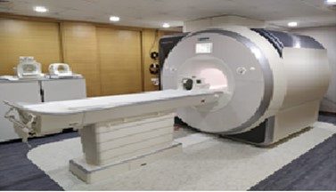
Neuroimaging is pivotal in understanding the structural and functional substrates involved in with healthy in brain aging and with various neurodegenerative and neuropsychiatric disorders. The Siemens PRISMA 3 Tesla fMRI machine has emerged as a game-changer for exploring the intricacies of the human brain. The Siemens PRISMA 3T fMRI machine housed at the J.N Tata MRI Centre in our campus and is a state-of-the-art magnetic resonance imaging system designed to provide unparalleled insights into brain structure and function. With its cutting-edge technology and advanced imaging capability, the PRISMA 3T is reshaping the landscape of pragmatic neuroimaging research and clinical applications.

This PRISMA fMRI machine capitalizes on a higher magnetic field strength, resulting in superior signal-to-noise ratio (SNR) with improved spatial and temporal resolutions. It empowers researchers at CBR & IISc to visualize fine details of brain structures and helps with accurately capture dynamic neural activity. The Siemens PRISMA incorporates TIM (Total Imaging Matrix) technology, advanced gradient system enabling high quality Diffusion imaging and tractographic studies and capable of parallel image acquisition. The Siemens PRISMA 3T fMRI machine also excels in mapping functional connectivity networks in the brain whereby our scientists leverage resting-state fMRI to investigate how different brain regions are regulated in healthy and pathological aging. The Siemens PRISMA 3 Tesla fMRI machine is a trailblazing innovation in the field of neuroimaging and an important player in CBR’s flagship efforts. With its advanced features, cutting-edge technology, unique and exceptional imaging capabilities, this imaging system enables researchers and clinicians to delve deeper into the mysteries of the human brain. By facilitating intricate studies of functional connectivity, diffusion patterns, and cognitive processes, the Siemens PRISMA 3T fMRI machine would therefore help to drive meaningful discoveries, shape clinical neuroscience research in India and pave the way for a comprehensive understanding of brain health and function.


Last Updated on September 29, 2023