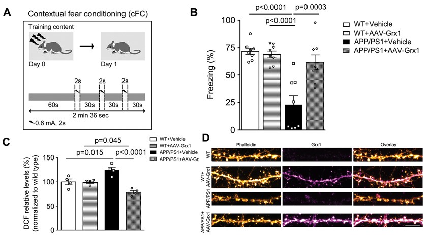
(A) Schematic representation of cFC experiment. (B) Bar graph representing freezing response in vehicle or AAV6-Grx1 injected WT or APP/PS1 mice. (C) Bar graph showing relative DCF levels in hippocampal lysates. (E) F-actin stabilization in AAV6-Grx1 expressing APP/PS1 primary cortical neurons.
Synaptic dysfunction, considered to occur early in AD is often seen as loss of dendritic spines, which are the sites of excitatory signalling in the brain. Fibrillar actin (F-actin) is highly enriched in dendritic spines and determines the shape of spine and undergoes restructuring during synaptic plasticity. We observed reduced levels of F-actin resulting in spine loss and memory deficits in AD mouse model. To understand the molecular mechanisms behind such reduction in F-actin in AD, we analysed the oxidative modifications of actin that affects its polymerization. We observed an increase in glutathionylation as a result of decreased levels of glutaredoxin1 (Grx1), an oxidoreductase enzyme that reduces glutathionylated proteins. This was associated with spine loss and memory deficits. Upon over expressing Grx1, we observed recovery in recall memory deficits, stabilization of F-actin levels and nanoarchitecture in dendritic spines. Our findings suggest that increasing Grx1 levels is a potential target for novel disease-modifying therapies for AD.
Last Updated on August 9, 2023
References :
Reddy Peera Kommaddi, Deepika Singh Tomar, Smitha Karunakaran, Deepti Bapat, Siddharth Nanguneri, Ajit Ray, Bernard L Schneider, Deepak Nair, Vijayalakshmi Ravindranath. Glutaredoxin1 Diminishes Amyloid Beta-Mediated Oxidation of F-Actin and Reverses Cognitive Deficits in an Alzheimer’s Disease Mouse Model. Antioxidants & Redox Signaling. 2019 Dec 20;31(18):1321-1338.
Reddy Peera Kommaddi, Deepika Singh Tomar, Smitha Karunakaran, Deepti Bapat, Siddharth Nanguneri, Ajit Ray, Bernard L Schneider, Deepak Nair, Vijayalakshmi Ravindranath. Glutaredoxin1 Diminishes Amyloid Beta-Mediated Oxidation of F-Actin and Reverses Cognitive Deficits in an Alzheimer’s Disease Mouse Model. Antioxidants & Redox Signaling. 2019 Dec 20;31(18):1321-1338.
![]()
